Phase contrast tomography at medium resolution

Phase contrast tomography at medium resolution was performed at the I13 beamline at the Diamond synchrotron radiation source (UK). The aim of the experiment was to probe sufficiently large volumes of lung tissue to reveal the spatial density and distribution of the asbestos bodies per unit volume. This information can be compared with the number of asbestos bodies per gram of dry tissue as calculated by optical and scanning electron microscopy. Several volumes of lung tissue samples from 4 human subjects with sizes of 2.1 x 1.8mm2 or 0.83 x 0.7mm2 (corresponding to resolutions of 0.8 and 0.33 micron, respectively) where probed.

A representative projection on the Z-axis of the tomographic data acquired is shown in the figure.

High resolution phase contrast and fluorescence tomography

Phase contrast and fluorescence tomographic data were acquired at the ID16A beamline at the European synchrotron on ~100 x 100 um2 samples (Figure 1). Besides the very small x-ray beam spot size achievable (down to ~10nm), which results in very high resolution, the great advantage of this beamline is the possibility to combine the information from fluorescence and phase-contrast tomographies on the very same sample. This eventually allows to obtain complementary information on the sample, such as 3D morphology and density (from phase contrast tomography) and 3D elemental distribution (from fluorescence tomography). In addition, the local density and thickness can be extracted from phase contrast data and used to calculate the absolute elemental concentration from fluorescence data. An example of a volume rendering of an asbestos body obtaine from phase contrast tomography is shown in the movie below.


Preliminary results from X-ray fluorescence mapping and X-ray near edge structure spectroscopy

Lung tissue of two former workers of an asbestos plant located in the Piedmont region (NW Italy), who have been subjected to prolonged exposure to crocidolite asbestos, was investigated using synchrotron radiation micro-probe tools.

Direct observation of the distribution of K and of elements heavier than Fe (Zn, Cu, As, and Ba) in the asbestos bodies (AB) was observed for the first time. In particular, Fe was found to be more concentrated in the inner area (Figure 1b), and P, K, and Ca were shown to be co-localized in the fibres’ coating (Figure 1a). The distribution of Si suggested the incipient dissolution of the inner fibre, and that of Zn, Cu, As, and Ba indicated that AB are efficient scavengers for these species, in agreement with the uptake ability of ferrihydrite, which is the mineral core of ferritin and hemosiderin.

Elemental quantification, also reported for the first time, confirmed that the coating is highly enriched in Fe (~20% w/w), while the other species are present in trace amounts (0.02 – 0.3 % w/w).

X-ray near edge absorption structure (XANES) spectroscopy indicated that Fe is in the 3+ oxidation state, and confirmed that it is present in the form of ferrihydrite, a poorly crystalline Fe oxide.

Comparison between the AB studied upon removing the biological tissue by chemical digestion with NaClO, and those embedded in histological sections, allowed unambiguously distinguishing the composition of the AB, and understanding to what extent the digestion procedure altered their chemical composition. In particular the comparison of the distributions of P, K, and Ca indicated that they are present as soluble species that are removed during the digestion of the tissue.

Iron enrichment in the inner part of the AB may be attributed to the gradual conversion of the primarily deposited and Fe-overloaded ferritin in the inner part of the AB into hemosiderin, with higher Fe content, and to subsequent precipitation of its ferrihydrite core. An alternative explanation may be the release of exogenous Fe from the asbestos fibre itself, which would undergo to gradual dissolution, as the observed distribution of Si suggested. The Fe distribution may also be determined by the combination of the above-mentioned mechanisms, one being a consequence of the other. In this framework a speculative model can be drawn in which the Fe-richer inner part of the AB develops with aging.

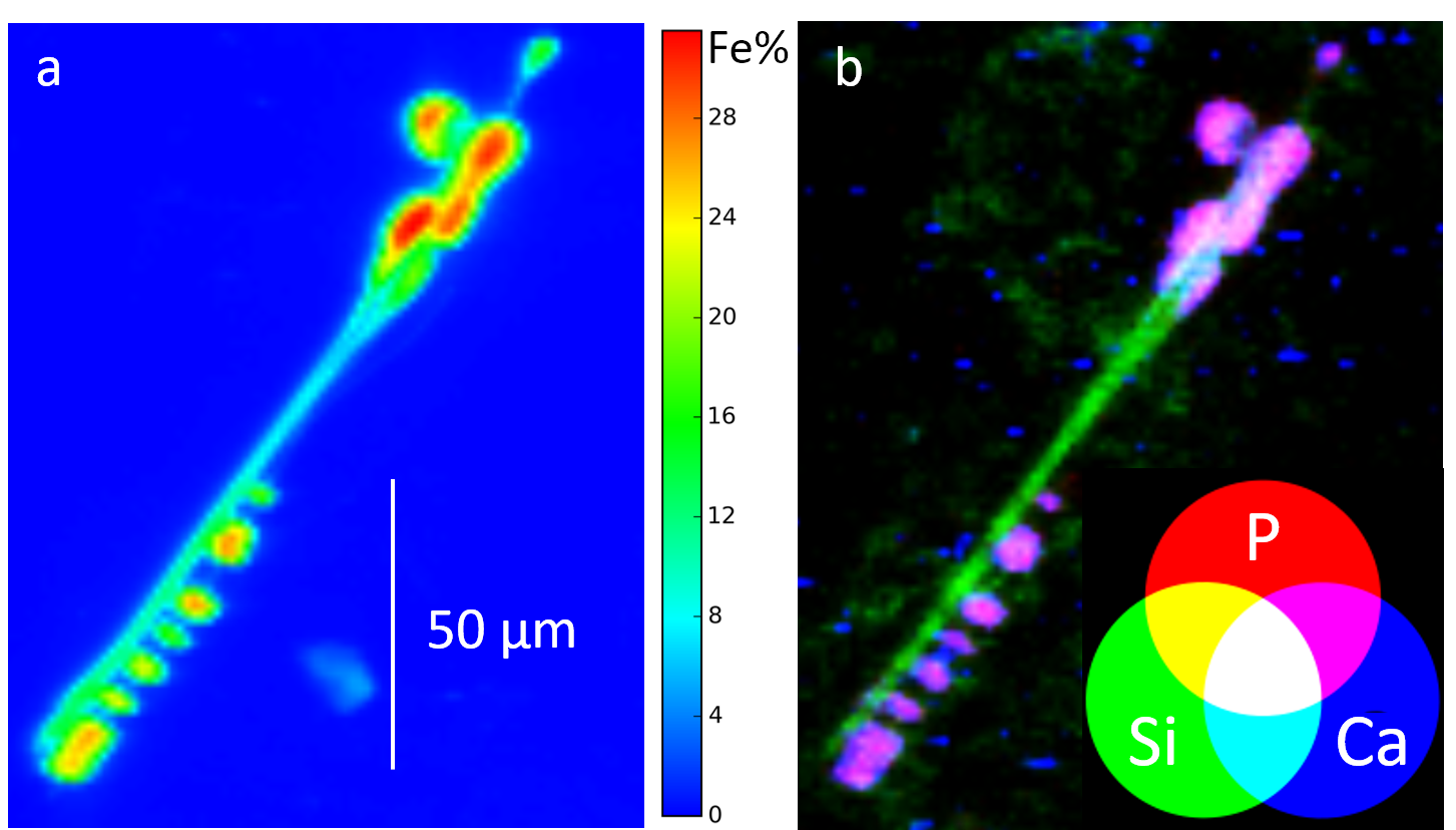
Figure 1. a) Distribution of Fe in an AB embedded in lung tissue. The color bar indicates the % w/w of Fe. b) Distributions of Si, P, and Ca in the same AB shown in a). The co-localization of Ca and P is evident. K has the same distribution of Ca and P (not shown). (Bardelli et al., 2017).

The work described above was published on the open access international journal Scientific Reports (Bardelli et al., 2017, doi: 10.1038/srep44862).