Samples

Human lung samples were collected after forensic autopsy from former workers of the asbestos plant in Cavagnolo, from a steel plant in Turin, and from the asbestos mine of Balangero. All of these activities were located in the Piedmont region (NW Italy) and are now dismissed. The cases were affected by pulmonary asbestosis, pleural plaques, pleural mesothelioma or lung cancer, and one had also silicatosis. The grade of asbestosis was established according to Craighead et al. (1982) [1]. Some of the Cases under investigation are summarized in the Table below. Other will be acquired and studied in the course of the project.
_____________________________________________________________________
|
Case
|
Age
|
Sex
|
Occupation
|
Exposure period
|
Exposure nature
|
Diagnosis
|
AB count (/g_dw*)
|
|
A
|
81
|
M
|
Asbestos Plant
|
27 years
|
Crocidolite
|
AS (grade 3) PP and MM
|
~3.6·10^5
|
|
B
|
80
|
F
|
Asbestos plant
|
unknown
|
Crocidolite
|
AS (grade 4) PP and LC
|
~1.2·10^7
|
|
C
|
80
|
M
|
Steel plant
|
unknown
|
Under investigation
|
AS (grade 4) MM, PP
|
~3.4·10^4
|
|
D
|
87
|
M
|
Asbestos mine
|
35
|
Chrisotyle
|
AS (grade 4) MM, PP, SS
|
~5.6·10^4
|
|
E
|
unknown
|
F
|
Textile Mill plant |
18
|
Under investigation |
AS MM, PP |
Under investigation |
|
_____________________________________________________________________ AB: Asbestos Bodies; AS: Asbestosis; PP: pleural plaques; MM: mesothelioma; LC: lung cancer; SS: silicatosis. *g_dw: grams of dry weight tissue |
|||||||
Samples' preparation
Lung samples were preserved in formalin (10%) until non-neoplastic portions (0.25 g) of lung tissue were digested in 30 mL sodium hypochlorite solution (NaClO, reagent grade, chlorine content 10-15%, Merck) to dissolve the organic matrix. The suspension of inorganic material was filtered through mixed cellulose esters porous membranes (pore size 0.45 µm, Millipore) to recover the asbestos bodies. The membranes were then thoroughly washed with warm (40°C) deionized water to dissolve NaCl crystals formed during digestion, and finally air-dried.
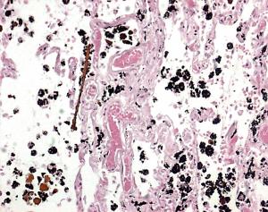
Further portions of non-neoplastic lung tissues were collected to prepare 3 micron thick histological sections with a microtome. The sections were embedded in paraffin, fixed on standard microscope slides, and then stained with haematoxylin and eosin, according to the standard protocol [2], for histological examination. Lung samples were examined to estimate the number of absestos bodies by optical microscope (Leica DMLB) and SEM (Cambridge Stereoscan S360). According to the international standard, the concentration of absestos bodies was expressed as their number per gram of dry weight (gdw). This quantity was derived following the procedure described in Belluso et al. (2006) [3]: (i) the whole membrane was observed by optical microscopy at 400x magnifications, and (ii) a portion of filter representing 0.7% of its total area (about 2 mm^2) was observed by SEM at 2000x magnifications. The equivalent dry weight was calculated by dehydrating 2.5g of wet tissue of each specimen at 60°C for 3 days. In both specimens the burden of AB largely exceeded the amount established by the European Respiratory Society guidelines (10^3/gdw) to indicate a high level of occupational exposure to asbestos [4].
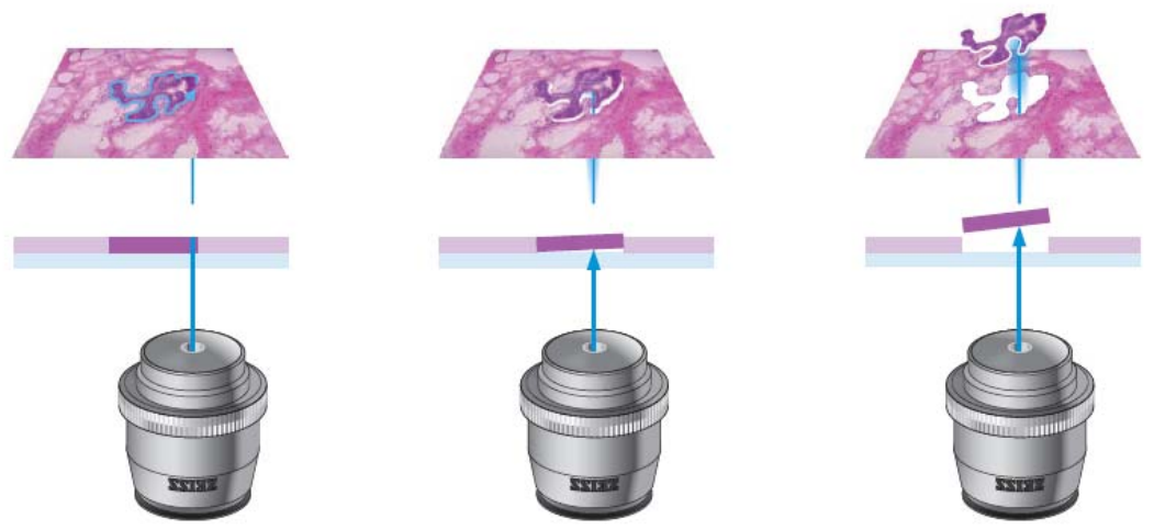
Due to the difficulty of locating the asbestos bodies in the histological sections, a laser micro-dissector was used to cut small areas (100 micron in diameter) of paraffin-embedded histological sections centred on the asbestos body. The fragments of lung tissue with one or more asbestos bodies inside were used for synchrotron tomography experiments. The principle of the laser-dissection procedure is described in the Figure below.
