Electron Microscopies
High resolution TEM images can reveal important information about the asbestos bodies surface, which being at the interface with the biological tissue, is expected to play a crucial role in the carcinogenesis. Electron diffraction, also available on transmission electron microscopes, could give complementary information on the crystallographic structure of the embedded asbestos fibres, supporting and complementing µXRD data. Transmission electron microscopy has not been widely used in the past for the present subject due to the lack of a reliable procedure to prepare the samples. This part of the project will focus on the development and test of such a procedure and on the acquisition of high quality TEM micrographs. The key feature of the proposed method is to minimize the steps needed to isolate and fix the fibers on the TEM sample holder, avoiding possible damage or artefacts induced by strong chemical attacks or mechanical stress. This goal will be obtained starting from the protocol developed by Cook [1] and combining mild digestion using sodium hypochlorite with several centrifugation steps. The minimal centrifugation time required will be determined theoretically by calculating the sedimentation rate of the fibrous material through a mathematical model.
[1] P M Cook. Preparation of extrapulmonary tissues and body fluids for quantitative TEM analysis of asbestos. Ann. New York Acad. Sci. 330 (1979) 717.

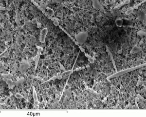
Scanning Electron Microscopy has been for decades the usual way to image asbestos bodies. It is an handy technique able to reveal their morphology with reasonable resolution, and also allows for semi-quantitative elemental quantification. Nevertheless, this latter information is limited to the surface (usually <1 micron), and, as for TEM, it is not possible (or very difficult) to image asbestos bodies embedded in the biological tissue. In this project SEM will be used to check the quality of the samples prepared for synchrotron radiation experiments, estimate the number of asbestos bodies per gram of dry tissue, and support SR-XRF quantification.